Variegated maple trees are uncommon but beautiful trees that have a white band
around the edge of their leaves. They are a form of Norway maple. Unfortunately,
the variegation is due to a virus, which can weaken the tree. The trees often
revert back to their natural green color. This particular tree had declined a
bit in past years, and this year had lost most of its leaves by the end of
August. So it was under more stress than usual. I cut off the tip of a branch
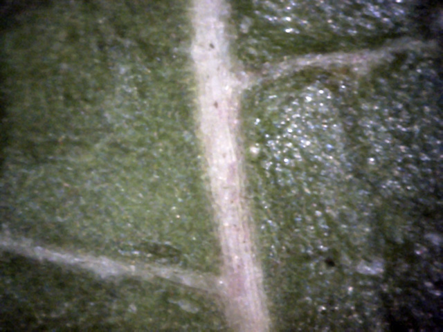
and checked it out, starting with the leaf top at 400x.
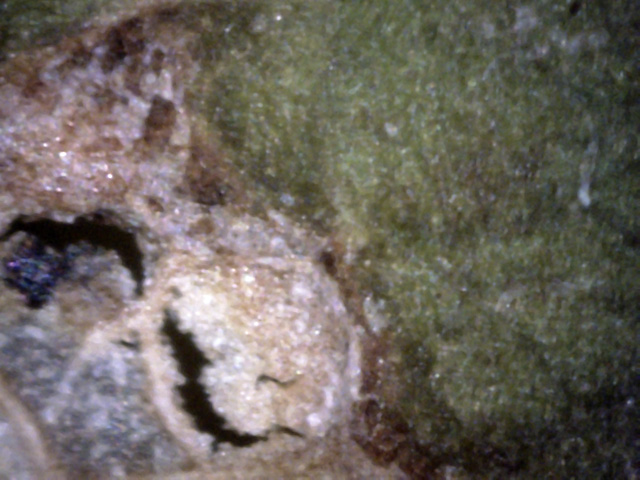
1
A dead area of the leaf. It looks like white canker may have killed it.
It also appears like a tiny strand of white canker is in the upper-right.
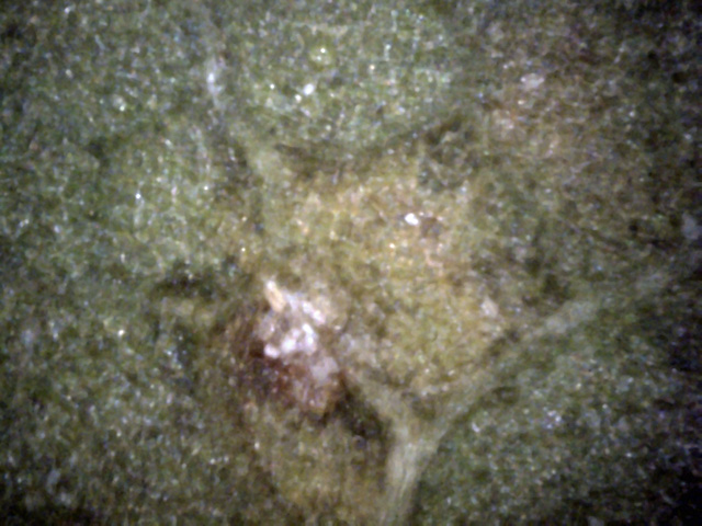
2
The white spots here appear to be a white canker infection.
Also, the leaf veins, which are mainly green, are starting to turn a bit white.
While leaf tops often show a diffuse growth and hint at internal canker growth, leaf bottoms are generally
a better indicator of white canker, as these 400x pictures show.
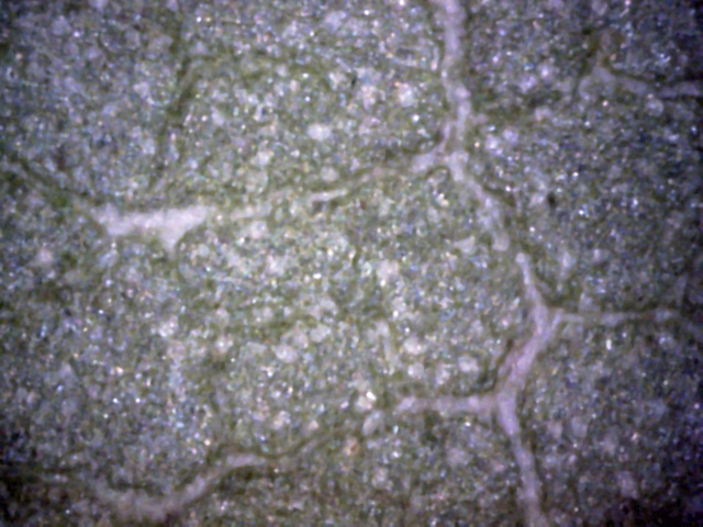
4
The leaf veins, which are normally green, are white here, indicating an infection.
Many little white white canker blobs are also seen.
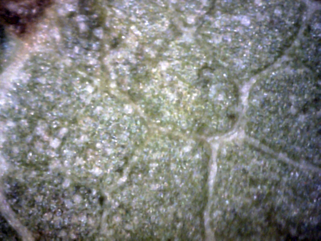
5
Here again, white leaf veins, and many white canker blobs, mainly close to the large leaf vein on the left.
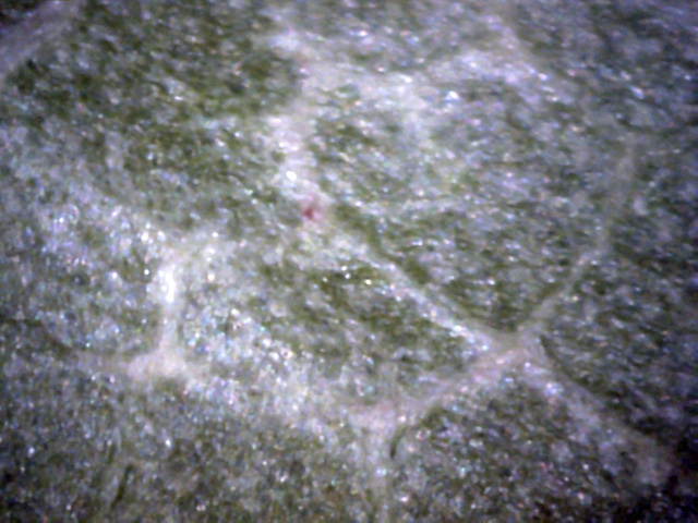
6
No white canker blobs here, but notice that the diffuse white growth seems to be located next
to the white leaf veins.
If white canker evidence is somewhat questionable when viewing the top or bottom of a leaf surface, a leaf cross-section
can settle the issue since it gives a view into the leaf, as shown in the 400x pictures below.

7
White canker material is visible on the top and bottom of this leaf.
White canker is also seen running from the top to bottom of the leaf on the left side of this picture.
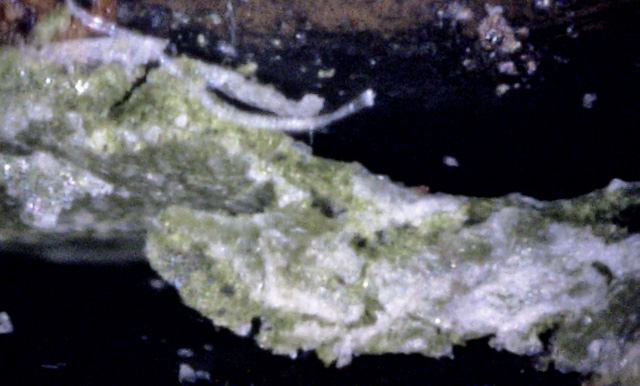 b
t
i
b
t
i
8
A leaf tear and cut make for an unusual view here, showing the leaf bottom (b), the top
(t), and the interior(i).
White canker particles can be seen on the bottom, while the white canker growth on the top is more diffuse.
There is little deep interior white canker growth - it all appears to be just touching the surface, much
as an iceberg.
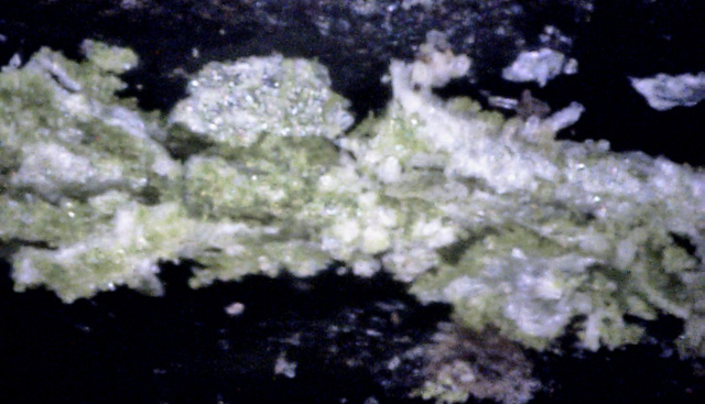
9
This part of the leaf is pretty torn up.
But notice the white canker particles in the middle of this picture and the diffuse growths to the right.
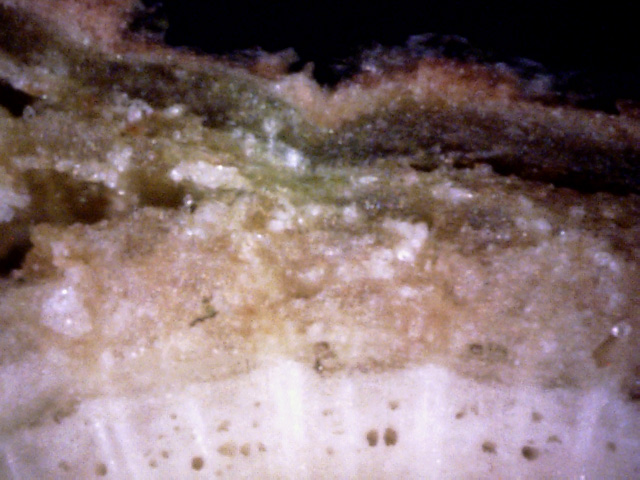
The diagnosis of white canker is most easily done by viewing a cross-section of a twig, as shown in the
400x microscope pictures below.
10
Extensive white canker damage is apparent. Many white canker particles are present.
The green phloem is almost totally gone, and has separated from the outer bark.
The normally somewhat green phloem appears to have been totally killed off and is evolving to a white color.
But the xylem still looks good, with open vessels, so the tree is getting water and minerals.
 ↓
↓
↓
↓
11
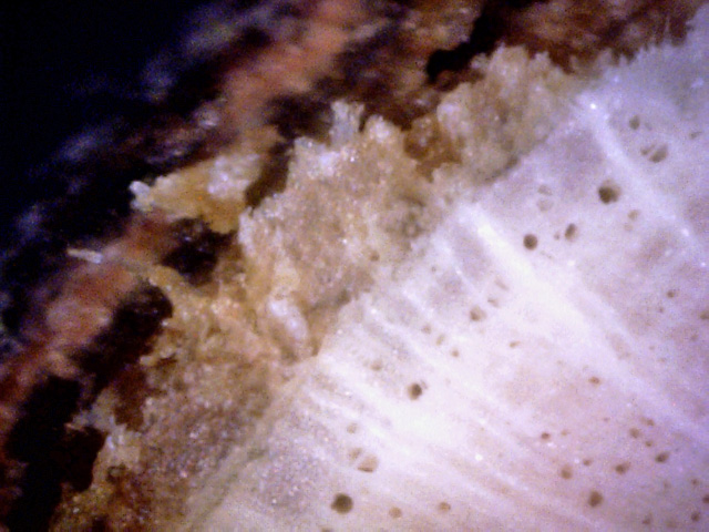
Notice that the cortex here is TOTALLY missing, having been replaced by an air gap.
The underlying phloem looks like it too will soon be gone.
Note the white canker growth starting in the phloem, going through the missing cortex, and then
penetrating the bark (red arrows).
12
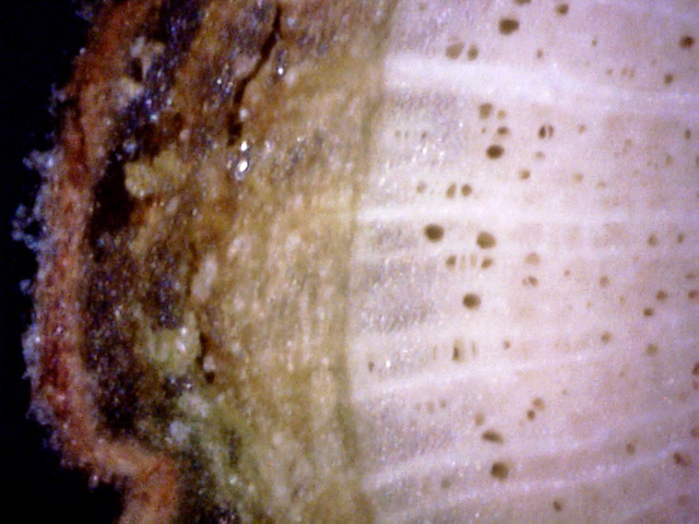
The good news is that the white xylem looks great!
The bad news is that the green phloem outside of it is half eaten and mangled by white canker.
Worse, the cortex is totally dead.
Finally, white canker growth is seen on the outside of the bark.
Strong confirming evidence of white canker is the presence of white canker spores on the bark.
But, you have to know where to look for them as they reside only in specific locations and are very
difficult to spot visually.
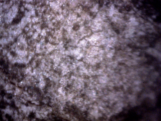
13
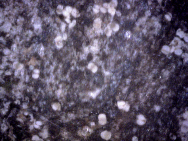
Much of the twig's outer bark surface looked like this - a light coating of white cankerous material,
much like a light snowfall.
14
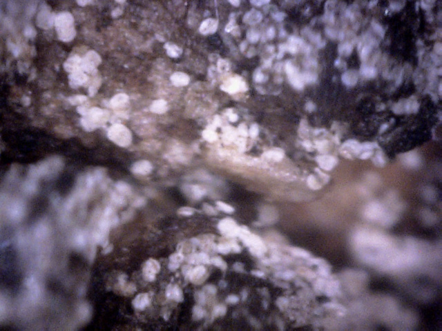
The final evidence of white canker: the particles themselves on the twig surface, having their very
characteristic spore shape.
Their embryonic yellow centers are still there, but muted due to age (the color saturation was boosted a bit
to more easily see them).
15
While 99% of the twig surface was free of spores, about 1% was loaded with them, and their location
was exactly as expected for a white canker infection - at twig junctions.
This is a view of one of those junctions.
The spore's bleached white appearance and brown centers imply that these spores are old - probably
3 months old.
In conclusion, the shriveled, brown, and dry appearance to the leaves of this tree gave an indication that
something serious was wrong. Other visual signs of white canker were not apparent. A microscopic examination
of the top, bottom, and cross-section of the leaves gave some supporting evidence of white canker.
Much stronger evidence came from a twig cross-section, which showed severe damage to the phloem and total
destruction of the cortex. The final piece of strong confirming evidence of white canker came from a microscopic
examination of the bark at a branch junction, where an abundance of white canker spores was seen.
